
Home > Conditions A-Z > not set


not set
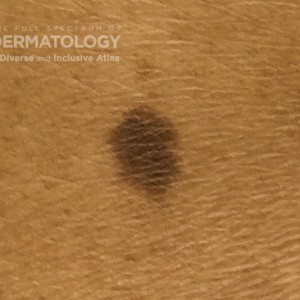
Nevi_Compound_B_Trunk_1.jpg
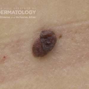
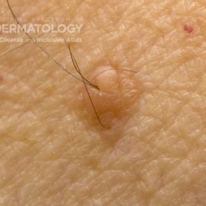
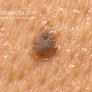
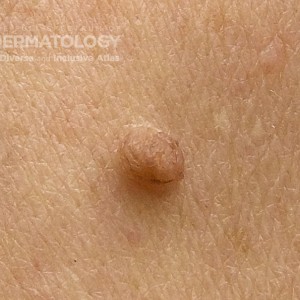
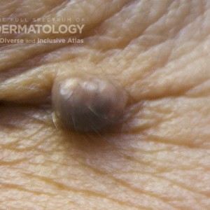
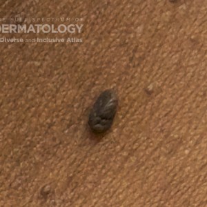
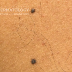
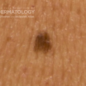
Nevi: Compound: These nevi may appear skin-colored to dark brown in lighter skin tones. In darker skin tones, they are often dark brown and can be distinguished from dermatofibromas by their fleshy to compressible architecture and by using dermoscopy.

Log into your JDD account to access high resolution images and request permissions.
Nevi_Compound_B_Trunk_1.jpg
https://cms.sanovaworks.com/uploads/2022/09/604fdacb60068cd5e2efa1d91c163eb0-small.jpg
Condition:
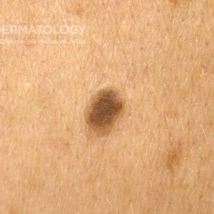
CompoundNevus_B_Chest1.jpg
Nevi: Compound: These nevi may appear skin-colored to dark brown in lighter skin tones. In darker skin tones, they are often dark brown and can be distinguished from dermatofibromas by their fleshy to compressible architecture and by using dermoscopy.

Log into your JDD account to access high resolution images and request permissions.
CompoundNevus_B_Chest1.jpg
https://cms.sanovaworks.com/uploads/2022/09/0eaf8c0645efe0826e7adda6aa3fb449-small.jpg
Condition:
NeviCompound_B_256.jpg
Nevi: Compound: These nevi may appear skin-colored to dark brown in lighter skin tones. In darker skin tones, they are often dark brown and can be distinguished from dermatofibromas by their fleshy to compressible architecture and by using dermoscopy.

Log into your JDD account to access high resolution images and request permissions.
NeviCompound_B_256.jpg
https://cms.sanovaworks.com/uploads/2022/09/43ae01cac4f750bf299a35ba0c5e1ef6-small.jpg
Condition:
CompoundNevus_A_Back1.jpg
Nevi: Compound: These nevi may appear skin-colored to dark brown in lighter skin tones. In darker skin tones, they are often dark brown and can be distinguished from dermatofibromas by their fleshy to compressible architecture and by using dermoscopy.

Log into your JDD account to access high resolution images and request permissions.
CompoundNevus_A_Back1.jpg
https://cms.sanovaworks.com/uploads/2022/09/5356394fc73334ac6ed8f54ee69de57c-small.jpg
Condition:
Nevi_Compound_C_Face_1.jpg
Nevi: Compound: These nevi may appear skin-colored to dark brown in lighter skin tones. In darker skin tones, they are often dark brown and can be distinguished from dermatofibromas by their fleshy to compressible architecture and by using dermoscopy.

Log into your JDD account to access high resolution images and request permissions.
Nevi_Compound_C_Face_1.jpg
https://cms.sanovaworks.com/uploads/2022/09/2bb2b7b4e40dc8a48b754958ef038989-small.jpg
Condition:
Compound_Nevus_D_Neck.jpg
Nevi: Compound: These nevi may appear skin-colored to dark brown in lighter skin tones. In darker skin tones, they are often dark brown and can be distinguished from dermatofibromas by their fleshy to compressible architecture and by using dermoscopy.

Log into your JDD account to access high resolution images and request permissions.
Compound_Nevus_D_Neck.jpg
https://cms.sanovaworks.com/uploads/2022/09/64ba4956a2ccebcba51a51d77cc9bc9a-small.jpg
Condition:
JunctionalNevus_A_Trunk_1.jpg
Nevi: Compound: These nevi may appear skin-colored to dark brown in lighter skin tones. In darker skin tones, they are often dark brown and can be distinguished from dermatofibromas by their fleshy to compressible architecture and by using dermoscopy.

Log into your JDD account to access high resolution images and request permissions.
JunctionalNevus_A_Trunk_1.jpg
https://cms.sanovaworks.com/uploads/2022/09/8ad784dd14d4e4ecc4dc41a43b3a4ae7-small.jpg
Condition:
JunctionalNevi_C_Chest3.jpg
Nevi: Compound: These nevi may appear skin-colored to dark brown in lighter skin tones. In darker skin tones, they are often dark brown and can be distinguished from dermatofibromas by their fleshy to compressible architecture and by using dermoscopy.

Log into your JDD account to access high resolution images and request permissions.
JunctionalNevi_C_Chest3.jpg
https://cms.sanovaworks.com/uploads/2022/09/27dcf7eade0f3a1d76b5cc642401f686-small.jpg
Condition:
NeviJunctional_B_256.jpg
Nevi: Compound: These nevi may appear skin-colored to dark brown in lighter skin tones. In darker skin tones, they are often dark brown and can be distinguished from dermatofibromas by their fleshy to compressible architecture and by using dermoscopy.

Log into your JDD account to access high resolution images and request permissions.
NeviJunctional_B_256.jpg
https://cms.sanovaworks.com/uploads/2022/09/633b55c14db4275530a8dedaf34492ba-small.jpg
Condition:
JunctionalNevus_E_leg2.jpg
Nevi: Compound: These nevi may appear skin-colored to dark brown in lighter skin tones. In darker skin tones, they are often dark brown and can be distinguished from dermatofibromas by their fleshy to compressible architecture and by using dermoscopy.

Log into your JDD account to access high resolution images and request permissions.
JunctionalNevus_E_leg2.jpg
https://cms.sanovaworks.com/uploads/2022/09/d66463eea52fc684c04ffe90f9c77031-small.jpg
Condition:
CONTACT US
We’re here to help and answer any questions you may have. We look forward to hearing from you!